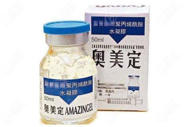
Many sisters mistakenly hit Omedine on the face (such as forehead, chin, nose), causing redness, swelling, pain, dissociation, deformation and other symptoms. They need to take it out immediately for health.
But, Omedine take out Before doing an accurate examination, in order to avoid residues, can the face be detected with customized CT, B-ultrasound and MRI by playing Ogilvy&Mather? Can you picture Omeidine? Today, I will reveal the answer to you.
Can the customized CT, B-ultrasound and MRI of facial Ogilvy&Mather take accurate pictures?
The answer is: Omedine can be seen through these three image methods, but compared with MRI, the ctb is not intuitive and clear enough.
Aili didn't help me make it up. It was verified by real people.
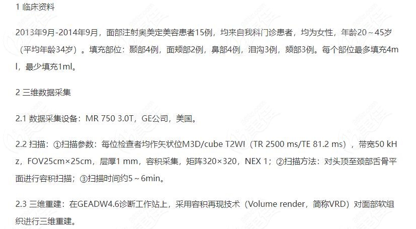
Note: Fifteen patients with facial injection of omedil were female, aged 20-45 years, (The average age is 34 years). Filling site: temporal region in 4 cases, cheek in 2 cases, nose in 4 cases, lacrimal groove in 3 cases, chin in 3 cases. More parts Charge 4m 1. Fill 1ml less.
There was a special MRI scanning and 3D reconstruction techniques were used to examine 15 cases of AUM fixed face syringes, The results show that The images taken by Meiding under MRI show clearly, which can more accurately assess the scope, boundary Residues and body morphology, Then, through one-time cleaning of the surgical incision, the purpose of less surgical trauma and greater surgical clarity can be achieved, and normal tissue damage can be avoided.
Take a look at the image of Omedin taken under the MRI:

Obviously, omedine is bright under NMR, and the white bright part is omedine. (Take the chest Olme fixed shot MRI as an example, the face Olme fixed shot MRI also shows white, very bright)
Come on Can CT Aomeiding take photos with B ultrasound?
The difference between CT B-scan and MRI is that B-scan uses sound waves to penetrate the face. If the strength is weak, it is difficult to detect small lesions. Although the CT image of Omedine is very clear, it has certain radiation. The resolution of MRI is high and the image is clearer. Even a small amount of Omedine can be photographed accurately, which is harmless to the body.
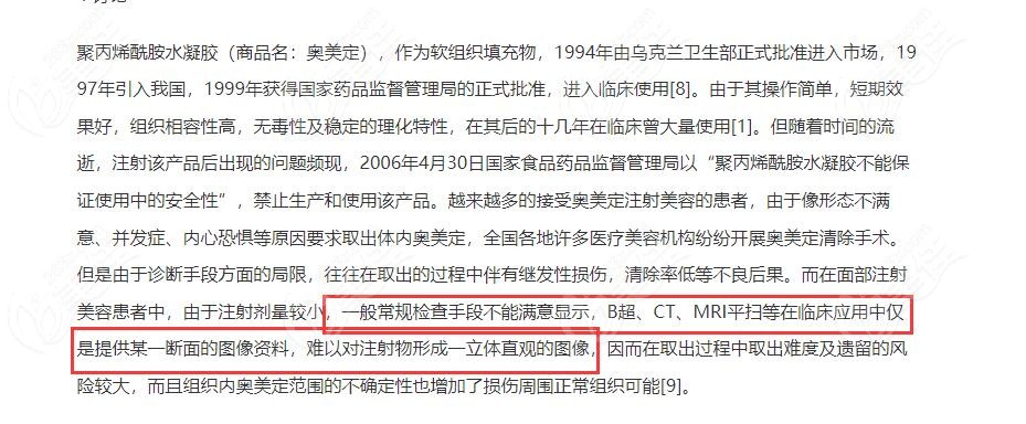
Example verification: general conventional inspection methods can not display satisfactorily, B ultrasound and CT only provide image data of certain sections, which is difficult to form a three-dimensional and intuitive image of the injection. It is difficult to take out during the removal process, and the residual risk is high z.com.
What kind of check does Choomey have to do? In general, MRI can accurately estimate the distribution level, scope, difficulty of surgery and degree of tissue damage of olmedine injection, so that it is cleaner to take it out at one time.
In other words, both the face and chest must be examined by MRI before removal, so as to more accurately see the distribution and scope of Ogilvy and improve the success rate of the operation.
Note: The diagnostic value of nuclear magnetic resonance and three-dimensional reconstruction in patients with facial filling in Austria and America, written by Shi Zehong, etc. Source: The 23rd issue of China Cosmetic Medicine in 2014.


